
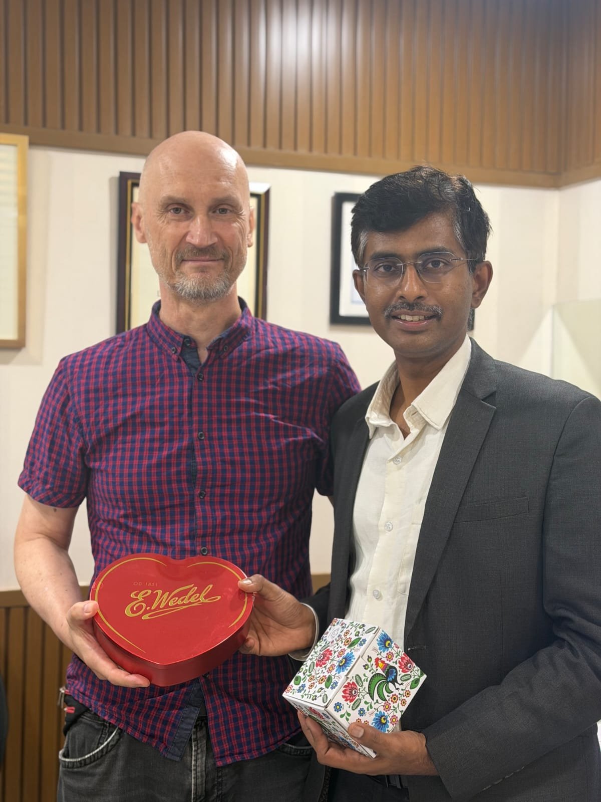
A 59-year-old man from Poland presented with recurrent palpitations and was found to have frequent ventricular premature complexes (PVCs). Following detailed electrophysiological evaluation, he successfully underwent radiofrequency (RF) catheter ablation for PVCs originating from the left ventricular summit.
Understanding Left Ventricular Summit PVCs
Ventricular premature complexes (PVCs), also known as ventricular premature beats or ventricular ectopics, are extra heartbeats that originate from the ventricles. While occasional PVCs may occur in healthy individuals, frequent or symptomatic PVCs can cause palpitations and may require further evaluation.
The left ventricular summit is a challenging anatomical region from which ventricular arrhythmias can originate. Due to its complex location and proximity to important coronary and cardiac structures, left ventricular summit PVC ablation requires detailed anatomical assessment and advanced electrophysiology techniques.
Clinical Presentation
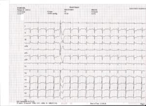
A 59-year-old male patient from Poland presented with recurrent episodes of palpitations. His initial electrocardiogram (ECG) demonstrated ventricular premature complexes, which were considered a likely explanation for his symptoms.
As part of the cardiac evaluation, the patient underwent a treadmill stress test, which was reported as positive. Considering the presence of symptomatic ventricular ectopy along with the stress-test findings, a detailed electrophysiology (EP) study was advised.
An EP study can help specialists identify the exact origin and electrical characteristics of abnormal heart rhythms and determine the most appropriate treatment strategy.
Electrophysiology Study and RF Ablation
The patient subsequently underwent an electrophysiology study and radiofrequency catheter ablation for PVCs arising from the left ventricular summit.
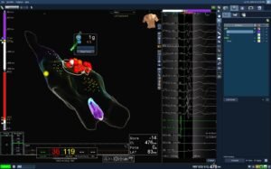
The procedure was performed with the support of advanced three-dimensional electroanatomical mapping and intracardiac echocardiography (ICE).
Three-dimensional electroanatomical mapping helps the electrophysiologist create a detailed representation of the heart’s anatomy and electrical activity. This allows the abnormal ventricular rhythm to be localised more precisely before delivering ablation therapy.
Intracardiac echocardiography provides real-time imaging from within the heart and can assist with anatomical localisation, catheter positioning and procedural safety.
Because of the complex anatomy of the left ventricular summit, combining advanced mapping with intracardiac imaging can be particularly valuable during complex PVC ablation procedures.
Successful Radiofrequency Ablation
After careful mapping and localisation of the PVC focus, radiofrequency energy was delivered to the targeted area.
The RF catheter ablation procedure was successfully completed, with successful treatment of the left ventricular summit PVCs.
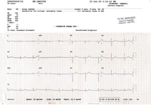
Following the procedure, the patient experienced symptomatic improvement. His post-procedure ECG demonstrated sinus rhythm within normal limits, indicating a favourable immediate response to the intervention.
The patient remained hemodynamically stable throughout the post-procedure period and was subsequently discharged with appropriate follow-up advice.
Why Accurate PVC Localisation Matters
The success of catheter ablation for ventricular arrhythmias depends significantly on identifying the precise site from which the abnormal electrical activity originates.
PVCs arising from challenging regions such as the left ventricular summit may require advanced electrophysiological mapping and careful assessment of surrounding cardiac anatomy.
Modern technologies such as:
- Three-dimensional electroanatomical mapping
- Intracardiac echocardiography (ICE)
- Advanced catheter navigation
- Radiofrequency catheter ablation
can support electrophysiologists in managing complex ventricular arrhythmias.
Takeaway from the Case
This case demonstrates the importance of a systematic approach to patients presenting with recurrent palpitations and symptomatic PVCs.
When ventricular ectopy is persistent, symptomatic, or associated with concerning clinical findings, a detailed cardiac and electrophysiological evaluation may help identify the underlying arrhythmia and guide treatment.
In selected patients, radiofrequency catheter ablation can provide an effective treatment option for symptomatic PVCs, including PVCs originating from anatomically challenging areas such as the left ventricular summit.
The successful outcome in this case highlights the role of advanced electrophysiology, three-dimensional mapping and intracardiac echocardiography in the management of complex ventricular arrhythmias.
About Dr. Deep Chandh Raja
Dr. Deep Chandh Raja is an electrophysiology specialist with expertise in the evaluation and management of cardiac rhythm disorders, including ventricular arrhythmias and complex catheter ablation procedures.
For patients experiencing recurrent palpitations, frequent PVCs, unexplained ventricular ectopy or suspected cardiac rhythm disorders, consultation with an experienced cardiac electrophysiologist can help determine the appropriate diagnostic and treatment pathway.